Nanoparticle Therapy – An Emerging Cancer Treatment
5 Min Read


The medical practice now uses artificial intelligence (AI) for automated diagnosis as this system learns to detect the patterns or signs of disease. For the identification of several ocular illnesses, such as diabetic retinopathy (DR), age-related macular degeneration (AMD), and glaucoma, AI systems are in use or under development.
The early types of medical AI were straightforward automated detectors that were taught to identify a predetermined set of disease-related characteristics. These early systems had the drawback of only recognizing patients who exhibit characteristics that are part of the specified program.
By examining a representative set of photos from people with and without the disease, and perhaps throughout multiple phases of the condition, the most sophisticated version of medical AI teaches itself to recognize the characteristics of sickness. The system goes through several iterations of analysis, assessment, and reanalysis during the learning phase until each picture can be accurately recognized. The number of illness characteristics that AI deep-learning systems may discover is unrestricted, in contrast to simple automated detectors.
Currently, automated AI systems are most frequently used in OCT. That’s what this page is dedicated to!
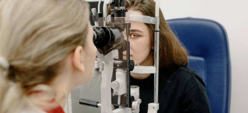
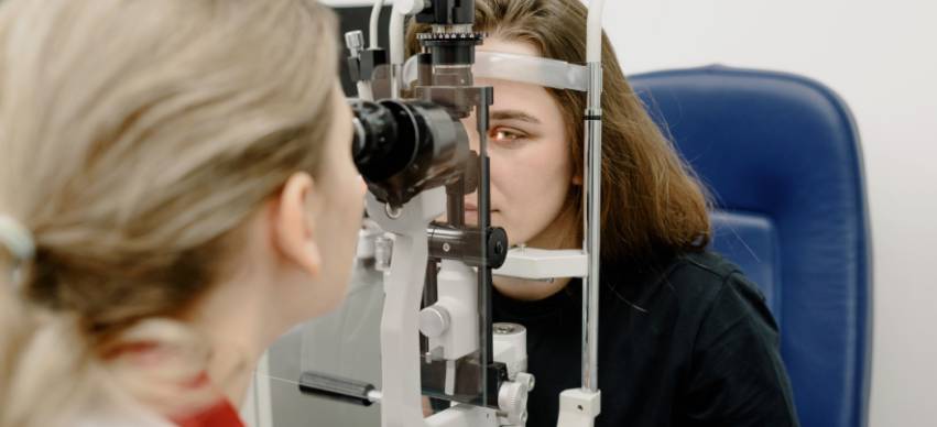
A non-contact, non-invasive imaging technique called optical coherence tomography (OCT) is used to see and track alterations in the shape of biological tissue. OCT uses low-coherence interferometry to provide cross-sectional pictures of the tissues of interest that show sub-surface features.
OCT devices produce high-resolution, volumetric pictures of tissue microstructures, including the cornea, iris, vitreous, crystalline lens, and retina, in the majority of typical ophthalmic applications. These photographs may improve understanding of pathological disorders like glaucoma, age-related macular degeneration, or diabetic retinopathy.
The benefits of OCT imaging include the next:
Ophthalmologists may expect new automated solutions from artificial intelligence (AI) for diagnosing and treating ocular diseases from the front to the back of the eye. This change is aided partly by the recent rise in interest from significant digital companies like Google and IBM in AI's potential for medical applications. However, according to ophthalmologists involved in these initiatives, computerized analytics are being looked at as a path toward more efficient and impartial ways to assess the stream of images produced by modern eye care systems.
Computer algorithms have the most potential in the field of retinal diseases. To give an example, there are companies that have already created AI OCT systems that are trained to identify a number of retinal conditions. These systems serve as the decision-making support for eye care specialists when dealing with complex OCT scans and early, rare, minor pathologies.
Beyond just the retina, artificial intelligence-based technologies are being developed to more accurately detect or analyze a variety of other ocular conditions, including juvenile cataracts, corneal ectasia, keratoconus, glaucoma, and oculoplastic reconstruction.
These algorithms can enhance objectivity in a range of circumstances, from screening to full management. Although doctors sometimes differ, an AI system always responds similarly and will even progress with time .
Multiple advantages of automated AI systems can boost healthcare practice. An AI system is, by definition, a tool for automated diagnosis, which can ease the load in a medical environment when physician resources are constrained. Data may be gathered remotely and transferred to an AI center for analysis since an AI system can execute its task anywhere. This is essentially an artificial intelligence (AI) version of telemedicine, where you can offer a diagnosis and other expert advice without the need for travel (patient or physician), increasing access to care for patients who live in remote or difficult-to-reach areas and easing the burden on patients, caregivers, and physicians.
A comprehensive series of closely spaced scans from a single patient that cover a vast area of the retina may be analyzed since AI systems can swiftly handle a lot of data. The likelihood of detecting early-stage illness rises as a result. Because treatment is frequently most effective when started early in a disease cycle before the tissue has been irreparably damaged, improving the capacity to detect early-stage illness is important.
AI systems may increase our understanding of illness processes and enhance therapeutic treatment. Deep learning AI has the ability to uncover hitherto undetected patterns of disease, enhancing ophthalmologists' comprehension of disease causation and adding new indicators for prognosis, staging, and diagnosis.